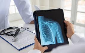
Artificial intelligence (AI) has transformed the medical industry in recent years, especially in the area of diagnostic imaging. AI-assisted bone fracture diagnosis in X-rays is among the most promising uses. This novel method improves accuracy, cuts down on diagnostic time, and helps physicians—particularly in high-stress settings like emergency rooms when accuracy and speed are essential.
AI in Radiology: The Basics
Radiology, the medical specialty that uses imaging to diagnose diseases, has seen tremendous growth in AI integration. AI systems, particularly those based on deep learning, are trained to analyze large datasets of medical images. These systems can recognize patterns and anomalies within the images, including fractures that might be missed by the human eye.
In the case of bone fractures, AI algorithms are fed thousands of X-ray images, both with and without fractures, allowing the machine to learn the subtle distinctions between normal bone structure and fractures. With this training, AI models can quickly and accurately assess new X-rays, flagging potential fractures for the clinician to review.
Improving Diagnostic Speed and Accuracy
One of the key advantages of using AI in fracture detection is the ability to enhance diagnostic speed. In a busy emergency room, doctors may need to review dozens of X-rays in a short time. Fatigue, stress, or complex cases can lead to missed fractures. AI helps mitigate this by serving as an additional layer of review. The AI system can quickly process the image, identify a potential fracture, and alert the clinician, allowing for faster treatment decisions.
Moreover, AI has been shown to improve accuracy. Studies have indicated that AI-assisted diagnosis can outperform human radiologists in certain scenarios, especially with subtle or complex fractures. While AI is not intended to replace radiologists, it acts as a powerful tool to augment their decision-making process, reducing human error and improving patient outcomes.
Applications and Real-world Use Cases
AI-assisted fracture detection is already being used in hospitals and clinics worldwide. Companies such as Zebra Medical Vision, Aidoc, and BoneView have developed AI software that integrates with existing radiology systems. These tools are especially useful in detecting fractures in high-risk patients, such as elderly individuals who may be prone to osteoporosis-related fractures or in trauma cases where multiple injuries need rapid attention.
In addition to detecting fractures, AI systems are also being trained to predict the severity of fractures, such as whether a fracture will require surgical intervention or if it can be treated conservatively. This predictive capability can help clinicians create more personalized treatment plans for patients.
Challenges and Future Prospects
Despite its potential, AI in radiology does face challenges. One of the major concerns is the risk of over-reliance on AI, where clinicians may become less vigilant in their review of medical images. To prevent this, AI should be used as an adjunct to human expertise rather than a replacement.
Another challenge is the variability in AI performance across different patient populations. AI systems trained on one demographic may not perform as well in another, making it essential to ensure that training datasets are diverse and representative of the broader population.
Looking ahead, the future of AI in radiology is bright. As technology continues to advance, AI models will become even more sophisticated, capable of detecting a wider range of conditions beyond bone fractures, such as infections, tumors, and degenerative diseases.
Wind-Up
AI is changing how doctors identify fractures on X-rays. AI is assisting in lowering the strain on medical professionals and enhancing patient care by speeding up and improving diagnoses. These systems have the potential to transform diagnostic imaging and guarantee that patients obtain prompt and precise diagnoses as they are more widely incorporated into daily practice. To optimize the advantages while lowering the hazards, cautious deployment and continual assessment will be necessary, as is the case with any new technology.


